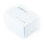
| Line | Method & Sample | Product | Package Info |
|---|---|---|---|
| MOLgen | Clinical Specimens | MOLgen Genetics AZF - Microdeletions Kit | Tests per Package: 48 |
| Genetics | Reagent kit is designed for differential determination of Y- chromosome AZF locus deletions by real-time PCR . The kits contain reagents for real-time PCR only. | Code: ME138140 | Package Format: STR |

For Quantity Orders: Request a Quote
Please pay attention to the revision of the document that must be the same as the revision reported in the box label.
In case of discrepancy please contact our Customer Care e-mail: info@adaltis.net.
* Other document related to the product available at Documentation Centre and it is accessible for Adaltis distributors/partners after registration only.
“MOLgen Genetics AZF-microdeletions Kit” is an assay kit for the differentialdetermination of Y-chromosome AZF locus deletions using real-time PCR.
“MOLgen Genetics AZF-microdeletions Kit” is intended for the differential determination of AZF locus deletions on the human Y-chromosome in clinical specimens (whole blood, buccal epithelium) using the method of real-time polymerase chain reaction (PCR) with fluorescence detection of amplified product.
The extraction of DNA from clinical specimens can be performed usingthe “MOLgen Universal Extraction Kit”manufactured by Adaltis S.r.l.
The results of PCR analysis are taken into account in complex diagnostics of disease.
The kitis intended for use with block-type PCR cyclers: AMPLIlab (Adaltis), CFX96 (Bio-Rad, USA), DT-96 (DNA-Technology, Russia).
The kit contains reagents required for 48 tests, including control samples.
NOTE:
The extraction protocol and the PCR set-up procedure can be done in automated mode using the Adaltis EXTRAlab Instrument by executing the extraction and PCR set-up protocol preloaded in the instrument user interface.
AZF microdeletions occur in the Y-chromosome regions responsible for spermatogenesis causing abnormalitiesin spermatogenesis and, therefore, male infertility. At molecular level, deletions extend over 0.8 - 7.7 Mb, including several genes, but the size of AZF loci is too small to be detected by cytogenetics methods, that is why “microdeletions” term is used. According to “EAA/EMQN best practice guidance for molecular diagnosis of Y-chromosomal microdeletions”, 2 specific marker sequences located in different regions of Y-chromosome AZF locus should be detected in order to confirm AZF microdeletion. The absence of any group of markers in the patient specimen DNA confirms the deletion of the corresponding AZF locus.
To detect AZFa, AZFb and AZFc deletions following specific marker sequences were selected: sY-84 and sY-86 (AZFa locus); sY-127 and sY-134 (AZFb locus); sY-254 and sY-255 (AZFc locus). The detection of SRY gene (that is responsible for the initiation of male sex determination in humans and is absent in females) is performed in order to confirm the presence of Y-chromosome DNA in the specimen and male gender confirmation. To confirm the presence of sufficient amount of human DNA in the specimen, the amplification of HMBS gene fragment located in autosome is performed simultaneously with Y-chromosome markers. The principal of detection of DNA marker sequences based on the amplification of the regions using the method of real-time polymerase chain reaction (PCR) with fluorescent detection of amplified product.
Real-time PCR is based on the detection of the fluorescence produced by a reporter molecule, which increases as the reaction proceeds. Reporter molecule is a dual-labeled DNA-probe that specifically binds to the target region of pathogens DNA. Fluorescence signal increases due to the separation of fluorescence dye and quencher by Taq DNA-polymerase exonuclease activity during amplification. PCR consists of repeated cycles: temperature denaturation of DNA, primer annealing and complementary chain synthesis.
Threshold cycle value (Ct) is a cycle number at which the fluorescence generated within a reaction crosses the threshold and the fluorescence signal rises significantly above the background. Increased signal is due to the use of a DNA hybridization probe that is specific for the given DNA sequence: it binds to one of the DNA strands in the course of reaction and provides additional specificity of the method. A DNA probe consists of a fluorescence dye at the 5’-end and a fluorescence quencher at the 3’-end that significantly reduces the fluorescence intensity. During the polymerase synthesis of the complementary strand, the probe is cleaved from the 5’-end due to the 5’-3’ nuclease activity of Taq DNA polymerase, the quencher and the dye become separated, thus increasing the fluorescence signal due to accumulation of the reaction product.
The detected fluorescence intensity depends on quantity of specific amplicons; the rate of fluorescence intensity increase depends on the quantity of human DNA in the specimen. To detect different amplicons, four fluorescent dyes (FAM, HEX, ROX, Cy5) are applied, reducing a number of tubes with reaction, thus making it possible to detect the products of different amplification reactions simultaneously in one tube.
The presence of a deletion in the locus is detected, if the amplification signal of a corresponding marker sequences is absent.
|
Reagent |
Content |
|
Control Sample (CS) |
1 vial, 1.0 mL |
|
Ready Master Mix for PCR No 1 (RMM 1)(lyophilized) |
48 tubes (tests) |
|
Ready Master Mix for PCR No 2 (RMM 2) (lyophilized) |
48 tubes (tests) |
|
PCR optical-quality film (or hinged caps) |
1.5 sheet (12 strips of 8 caps) |
|
Number of tests |
48 |
|
Code |
ME138140 |
Need assistance to make an order? Contact Sales & Orders Centre order@adaltis.net.
For Application Support application@adaltis.net